From cellular structures and microbial morphology to the microstructure of materials and crystal defect analysis, microscopes play a critical role in scientific exploration, medical diagnostics, and industrial inspection. With their unique capability to reveal the unseen, microscopes have become indispensable tools across many fields. In this blog, we’ll take a comprehensive look at the definition, development, working principles, main types, and applications of microscopes—your precision eye into the microscopic world.
Definition and Nature of a Microscope
A microscope is an optical instrument designed to magnify tiny objects or microscopic structures. Its core function is to render images of objects too small to be seen by the naked eye, using optical or electron optical systems. The magnification of a microscope often far exceeds human vision (which has a resolution of about 0.1 mm), allowing objects to be enlarged hundreds, thousands, or even millions of times, revealing intricate micro-level 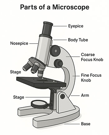 details.
At its core, a microscope is a sophisticated integration of optics, mechanics, and electronics—a truly mechatronic instrument. It combines precise optical system design, high-precision mechanical engineering, and advanced electronic control to achieve high-resolution imaging. Key performance indicators of a microscope—such as magnification, resolution, depth of field, and working distance—directly determine its effectiveness and range of applications.
Historical Development of Microscopes
The history of the microscope dates back to the early 17th century and has evolved from simple optical microscopes to modern electron microscopes.
- The Birth of Early Optical Microscopes: In 1590, Dutch spectacle makers Hans Janssen and his son Zacharias Janssen are credited with inventing the first compound microscope by combining two convex lenses to magnify distant objects. However, these early optical microscopes had limited magnification and image quality due to basic optical technology. In 1632, Dutch scientist Antonie Van Leeuwenhoek significantly improved microscope design using a single-lens (simple microscope) with finely polished glass spheres as the objective lens. His microscope achieved up to 270X magnification and allowed him to observe microorganisms like bacteria and protozoa for the first time, paving the way for microbiology.
- Advancements in Modern Optical Microscopes: By the mid-19th century, with progress in optical theory and glass manufacturing, the performance of optical microscopes greatly improved. German scientists Ernst Abbe and Carl Zeiss developed the Abbe sine condition, which became a theoretical foundation for microscope optics. Zeiss also introduced achromatic and apochromatic objectives, significantly enhancing resolution and image clarity in modern optical microscopes.
Basic Principles of Microscopy
The working principle of a microscope is based on optical or electron optical imaging, using a combination of objective lenses and eyepieces (or electron optics) to magnify microscopic objects.
- Imaging Principle of Optical Microscopes: An optical microscope uses visible light as the illumination source. The objective lens collects light from the specimen to form a real image, which is then magnified again by the eyepiece into a virtual image visible to the human eye. Following geometric optics principles, total magnification equals the product of the objective and eyepiece magnifications. The resolution is limited by the wavelength of visible light; according to Abbe's diffraction limit, the theoretical resolution is about half the wavelength, roughly 200 nanometers.
- Imaging Principle of Electron Microscopes: An electron microscope uses an electron beam instead of light, and since electron wavelengths are much shorter than visible light, electron microscopes offer much higher resolution. Transmission Electron Microscopes (TEM) transmit electrons through the specimen to form images, while Scanning Electron Microscopes (SEM) scan the specimen surface, detecting secondary electrons to create surface morphology images. Electron microscopes can achieve atomic-level resolution and are vital tools for studying material microstructures and crystal defects.
Main Types of Microscopes
With continuous technological innovation, various types of microscopes have emerged, each with unique imaging principles, structural designs, and application fields. Below, we introduce three representative microscope types offered by SISCO: digital microscopes, biological microscopes, and fluorescence microscopes.
1. Digital Microscopes: A digital microscope is a modern integration of traditional optical microscopy and digital imaging technology. It uses a built-in high-resolution digital camera to capture images in real-time, converting them into digital signals for display, storage, and analysis on computers or other devices. Key advantages of digital microscopes include.
- Real-Time Display & Collaboration: Digital microscopes display live images on a screen, enabling multiple users to observe and discuss simultaneously—ideal for microscope education and research collaboration.
- Image Processing & Analysis: Specialized software allows users to adjust brightness, contrast, color balance, perform measurements, annotations, stitching, and more, making it a powerful microscope for research.
- Data Storage & Review: Captured images and videos can be stored for future analysis, aiding in scientific documentation and teaching materials.
- Remote Operation & Monitoring: With network connectivity, digital microscopes support remote viewing and control, improving flexibility and efficiency.
Digital microscopes are widely used in education, scientific research, industrial inspection, and medical diagnostics. They help students explore the micro-world, enable researchers to conduct precise analysis, support quality control in manufacturing, and assist in pathology diagnostics.
2. Biological Microscopes: A biological microscope is designed specifically for observing biological specimens and is one of the most commonly used tools in biological research and teaching. It typically uses transmitted light to display cell structures and tissue morphology. Key structural features include.
- Advanced Optical System: Equipped with high-resolution optics and multiple objective lenses for selecting suitable magnification levels.
- Specimen Stage: Precisely adjustable platform for positioning biological samples, enabling targeted observation.
- Focusing Mechanism: Both coarse and fine focus knobs allow for sharp focusing, accommodating samples of varying thickness.
Biological microscopes are extensively used in biology, medicine, and agriculture. In biology, they help visualize cell structure and developmental processes; in medicine, they assist in pathology, blood analysis, and microbiology; in agriculture, they support plant disease diagnosis and seed quality inspection. If you're looking for a microscope for biological samples, this type is ideal.

3. Fluorescence Microscopes: A fluorescence microscope utilizes fluorescence to form images. It excites fluorescent substances in the specimen using specific wavelengths of light, which then emit light of a longer wavelength (fluorescence). This fluorescence is captured and magnified by the optical system to form an image. Typically, high-pressure mercury lamps or lasers are used as light sources. Once the sample absorbs the excitation energy, it emits fluorescence as it returns to its ground state. Filter sets selectively transmit fluorescence while blocking excitation and stray light. Advantages include.
- High Specificity: Fluorescence microscopes can label and visualize specific biological components using targeted fluorescent dyes.
- High Sensitivity: Fluorescent signals stand out against complex backgrounds, enhancing detection accuracy.
- Multicolor Imaging: By applying multiple dyes, users can observe several components simultaneously, making this ideal for studying molecular interactions and spatial distributions.
Fluorescence microscopes are widely used in cell biology, molecular biology, and biomedical research. They help track protein distribution, signal transduction, and gene expression, and are instrumental in disease diagnostics like tumor marker detection and virus identification.
Conclusion
As a precision tool for exploring the microscopic world, the microscope has evolved alongside human understanding of nature. From early optical microscopes to today’s high-resolution electron microscopes—and the innovation of digital, biological, and fluorescence models—microscope technology continues to push the frontiers of science.
Looking forward, as fields like nanotechnology and quantum science progress, SISCO believes that microscopes will play an even more vital role in uncovering the mysteries of the microscopic world. Whether in biomedicine, materials science, or industrial production, the microscope remains a cornerstone of scientific advancement and human development.